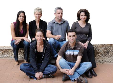
Our immune systems are hugely complex cellular networks that depend on each member carrying out its task. Dr. Guy Shakhar of the Immunology Department is a sort of immune system sleuth, staking out immune system cells and tailing them to find out where they go, how they get there and who they talk to. His surveillance equipment consists of a sophisticated imaging microscope (see box) that enables him to observe these cells both in their normal daily routines and in the act of abetting disease.
One group of cells that Shakhar and his team track is dendritic cells. These cells hang around in organs like the gut, skin and lungs – places where pathogens like to enter the body. Dendritic cells sample any foreign matter they find and, if further action is called for, whisk their sample – a bit of protein from the pathogen called an antigen – off to the nearest lymph node to present it to other cells of the immune system. These are primarily T cells that can kick-start an immune response, kill dangerous cells and recruit other immune cells – such as the B cells that produce disease-fighting antibodies. Shakhar is investigating how dendritic cells mobilize, traveling from the skin or gut through the lymph ducts and into the lymph nodes. It's known that these cells don't travel well if their surface receptors for two molecules – one that allows them to sense their target and another that helps them adhere – are blocked. Shakhar is trying to find out exactly when these molecules come into play – whether they help the immune cells leave their patrol sites, steer them to their destinations or enable them to enter the lymph nodes.
But sometimes the pathogen invasion is more of a sneak attack: For instance, just a few malaria parasites in a mosquito's bite are enough to cause disease. If these parasites are unfamiliar to the immune system, only a few out of the billions of patrolling T cells may have surface molecules that can identify the new antigens. A race begins between pathogens and immune system, and this small number of cells must convince the immune system to launch a full-scale attack on short notice. The team's evidence suggests that the T cells and dendritic cells combine forces, the dendritic cells forming networks that coordinate their presentation. The T cells can then navigate these networks to find the most informative antigen-presenting dendritic cells. The team is currently exploring how this coordination is carried out.
Shakhar is also on the trail of a network of cells involved in a family of lower digestive system disorders that includes Crohn's disease. Evidence suggests these autoimmune diseases are rooted in the faulty regulation of immune responses to the normal population of microbes living in the intestines. This network involves three-way interactions between the inflammation-causing immune system cells, regulatory T cells, which damp down immune responses, and dendritic cells. He and his team are imaging the intestines of mice into which they have introduced both bacteria with specific antigens and immune system T cells that recognize and respond only to these antigens. They believe the dendritic cells are key players in this network, and the team's efforts to pin down their role might provide new targets for treating these diseases.
True to Life
When Robert Hooke first observed a slice of cork under a microscope in the 17th century, he named the tiny empty compartments he saw "cells." In the ensuing years, microscopes have become much more powerful, revealing the world of cells in infinitely greater detail. But until recently, whenever researchers wanted to visualize cells inside complex tissues, they had to fix them firmly in place – as dead as those in Hooke's cork.
In the last few years, new kinds of microscopes have enabled scientists to observe living, moving cells deep inside live animals.
Dr. Guy Shakhar uses one called a two-photon microscope to track immune cells in anesthetized mice. Based on a physical phenomenon in which two infrared photons striking fluorescent molecules in rapid succession cause them to emit a flash of visible light, the method produces 3-D images of cells and tissues by shining ultra-quick pulses of highly focused infrared laser light. The infrared beam can penetrate tissue to a depth of several hundred microns, allowing Shakhar to observe the organization of the tissue without slicing into it and to follow the activities of cells over time.
Image of a Scientist
Dr. Guy Shakhar was born in Jerusalem and grew up in Givatayim, near Tel Aviv. After serving in the IDF, he enrolled in Tel Aviv University, receiving an M.Sc. in interdisciplinary studies and a Ph.D. in neuroscience, in which he researched interactions between the immune system and the nervous system. In his postdoctoral work with Prof. Michael Dustin at NYU School of Medicine, Shakhar turned to imaging immune cells to investigate their activities. In 2006, Shakhar joined the Weizmann Institute as a senior scientist.
Married to Keren, Shakhar is the father of two children – Amit, aged four and Gal, aged six. He enjoys mountain biking and playing the Asian board game Go.
Dr. Guy Shakhar's research is supported by the Kirk Center for Childhood Cancer and Immunological Disorders; the Crown Endowment Fund for Immunological Research; the Linda Tallen and David Paul Kane Educational and Research Foundation; the Philip M. Klutznick Fund for Research; the Wolfson Family Charitable Trust; and the estate of David Turner.
