Even in the most thoroughly researched fields, surprises can still turn up. Once in a while these surprises challenge the accepted wisdom, even when that wisdom is well founded in fact. For instance, scientists studying the family of enzymes known as proteases recently found a new type of protease that seemed to stand the body of knowledge about these enzymes’ actions on its head.
Proteases (literally: protein cutters) are a large collection of enzymes responsible for clipping proteins. Because they are found in every life form and play key roles in numerous basic life processes, proteases are some of the best-studied enzymes around. But recent research turned up evidence of another long-lost branch of the family. While their well-known cousins are to be found floating in fluids inside or around the cell, these proteases are hidden away, embedded in the cell membrane. It was the enzymes’ location inside the membrane that created a paradox for the researchers: The proteases they had studied require water molecules to help them cleave proteins. The interior of the cell is hydrophilic – water-loving – and the enzymes have no trouble finding water molecules to use there. By contrast, the cell’s outer wall – the cell membrane – is made of fatty molecules that are water-repellent. How, then, could a protease function in this environment?
At first, scientists, steeped in the common wisdom, doubted the evidence hinting at the existence of membrane-bound proteases. Findings, however, continued to mount showing that they not only exist, but are involved in a range of important activities in the cell. These activities include intercellular communication, signaling inside the cell, regulating programmed cell suicide and preventing invasion by parasites. Membrane proteases also play a role in the formation of beta-amyloid protein segments such as those that accumulate in the brain in Alzheimer’s disease.
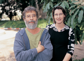
Yet the central question re-mained: How does protease activity, dependent as it is on water, take place in a water-repellent environment such as the cell membrane? To answer this question, Prof. Eitan Bibi and postdoctoral fellow Dr. Adam Ben-Shem of the Biological Chemistry Department, together with Prof. Deborah Fass of the Structural Biology Department, succeeded in solving the three-dimensional structure of a protease found in the cell membrane of the bacterium E. coli. The study, which recently appeared in the Proceedings of the National Academy of Sciences (PNAS), points to a possible mode of action for this enzyme – one that suggests an answer to the paradox.
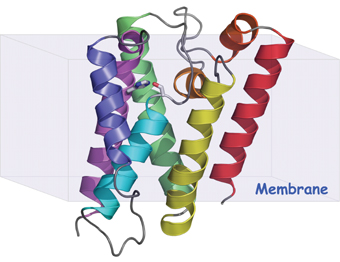
The enzyme is made up of six coils joined together by loops. Five of the six coils form a sort of cylinder that transverses the membrane, extending out past the membrane surface on either side. Inside this tube, the sixth coil, shorter than the others, harbors the protease’s active site – where the actual cutting is performed. Situated right above the active site, supported by the ring of coils, is a sac-like structure that’s padded with amino acids. These amino acids carry electrical charges on their ends that attract water, and the sac’s position implies that the water molecules are then funneled down to the active site to create a hydrophilic mini-environment in which the enzyme can work.
In solving one mystery, however, the scientists stumbled on another: How does the protein that’s cut by the protease gain access to the active site, which is set deep within the enzyme structure and surrounded by closely packed protein coils? The research team’s findings suggest a number of possibilities. In one place, the structure showed evidence that one of the loops binding two of the outer coils together might act as a gate that opens to let in the protein molecule. Alternately, a V-shaped opening that showed up between another two coils might give the protein access. But both openings are far from the active site, and the scientists believe that protein, protease or both probably need to undergo a change in shape for cleavage to take place. “A change in the structure of the substrate protein might allow it to access the enzyme’s active site and also expose the spot that needs cutting,” says Bibi.
The next stage of inquiry poses a challenge to the researchers. The enzyme’s location makes it hard to study, and they are currently searching for ways to observe the changes it undergoes as it cuts proteins deep inside the cell membrane. Because membrane proteases are so widespread and are vital to so many of the cell’s functions, this research is likely to have an impact on a wide range of biological and medical research.
Prof. Eitan Bibi’s research is supported by La Fondation Raphael et Regina Levy.
Prof. Deborah Fass’s research is supported by the Clore Center for Biological Physics; and the Helen and Milton A. Kimmelman Center for Biomolecular Structure and Assembly.


