When a tumor is discovered, the first question asked is: malignant or benign? The usual method for making a determination is to do a biopsy. This can be painful and even disfiguring. And even when the news is good, it's a tough way to find out. All this may soon change.
A non-invasive alternative for discovering and distinguishing between tumors using magnetic resonance imaging (MRI) has been developed by the Weizmann Institute's Prof. Hadassa Degani, who is now successfully applying it to breast cancer in humans.
The technique, featured recently on the cover of Nature Medicine, can reveal tumors as small as one cubic millimeter, denote their nature and even indicate how aggressive a malignant tumor may be. Degani's method promises to allow doctors to make more accurate diagnoses and monitor and adjust therapies accordingly.
According to Degani, a member of the Biological Regulation Department, "Most breast tumors detected by mammography are revealed to be benign on biopsy, so a non-invasive method like MRI could help reduce the rate of unnecessary procedures."
With Degani's technique, patients are spared potentially harmful X-ray radiation or surgery. A dye-like contrast substance is injected into the bloodstream and is tracked as it moves into and out of the tumor and its surrounding tissue. The MRI image is built up using a technique Degani developed called 3TP (Three Time Point). In 3TP, a "snapshot" of the breast is made once before the dye is injected and twice more at intervals of several minutes. Recording each image takes from two to four minutes, instead of the usual several seconds. Unlike earlier attempts at MRI imaging of tumors, Degani's method provides high-resolution pictures on a computer screen, with benign and malignant tumors showing up in different colors.
In developing her revolutionary new method, Degani exploited the different properties of malignant and benign growths. In malignant tumors, the cells are densely packed with very little intercellular space between them, and they are fed by many small blood vessels that are porous and leaky. Benign tumors, on the other hand, are less densely packed and have fewer blood vessels.
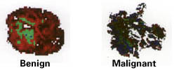
After the dyes are absorbed, the cells reveal these physiological differences in color: red in areas of minimal leakage, green in areas with steady levels, and blue where the leakage is rapid. A benign tumor, having more space between cells and containing fewer blood vessels, takes up and releases the contrast substance slowly. In the denser malignant tissue, the dye passes through the tissue quickly and does not accumulate. The malignancy's many blood vessels are also more porous, leaking the color into the intercellular space. These traits make the 3TP images clearly definable as benign or cancerous tumors, and give new meaning to the phrase in living color.
Degani's team looked at 18 cases, eight of them fibroadenomas (benign tumors) and 10 of them breast cancers. The fibroadenomas looked mostly red with patches of green. In the cancerous tumors, blue predominated. Furthermore, the colors in the benign tumors were uniform and well-defined, denoting slow uptake, accumulation and wash-out. The malignant tumors showed colors distributed in chaotic, uneven patches, indicating wild and rapid processing.
The next step is large-scale clinical trials. If the results are consistent with the early findings, Degani's 3TP approach may become the tool of choice for detection and diagnosis of cancer in the breast and other organs.
This project was funded in part by the U.S. National Cancer Institute and the National Institutes of Health; the Israel Academy of Sciences and Humanities; the German-Israeli Foundation for Scientific Research and Development; and the Weizmann Institutes Canadian Women for Science.
