About one in 4000 babies is born with DiGeorge syndrome, a congenital condition that causes various abnormalities, most often in the face and heart. This syndrome usually arises from the deletion of a small portion of chromosome 22, but strangely enough, the symptoms and severity vary greatly among individuals. Facial defects can range from cleft palate or difficulty in eating or speaking, to mild muscle weakness on one side of the mouth; and heart defects can include deformities in the wall separating the chambers or misplaced aorta.
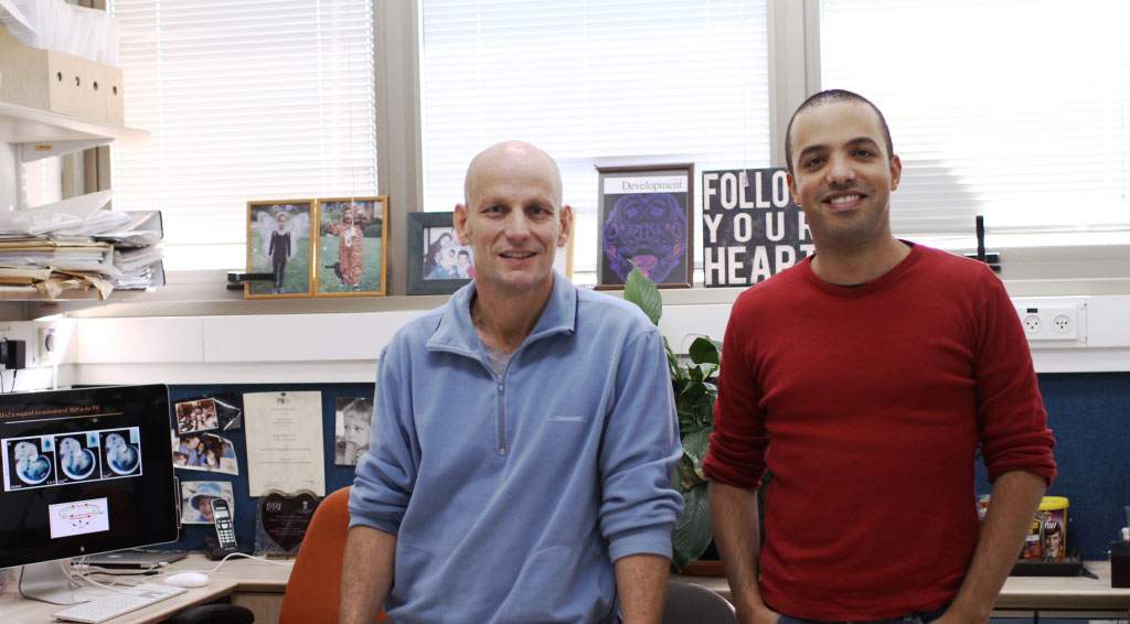
Prof. Eldad Tzahor and research student Itamar Harel of the Institute’s Biological Regulation Department recently solved a piece of this puzzle by investigating the genetic network underlying this syndrome.
Their findings appeared in the
Proceedings of the National Academy of Sciences (PNAS).What genes are important for proper heart and head muscle development? Is more than one gene involved and, if so, how do they function together? Tzahor and Harel focused on transcription factors – proteins that bind to DNA and control the transcription (the first step in converting genetic information to proteins) of other genes. An initial screen for the factors that are active in these early mesoderm cells turned up some that were already associated with the development of these cells, as well as a new one called Lhx2. This transcription factor was known to be involved in other areas of development – including eyes, blood cells and hair follicles – but it had never been seen to play a role in heart or muscle development.
Next, the researchers looked for the effects of these transcription factors in living organisms. In some of their mice, each of the main transcription factors they had identified was individually knocked out. But they also managed to create several “double knockout mice,” in which two of the genes were inactivated. When just one gene was knocked out, the team observed various DiGeorge-like deformities in the heart and facial muscles – some mild and some more severe. But when two were knocked out at the same time, the embryonic cells failed to “graduate to second grade,” and specific muscles and heart structures did not form.
Combining these results with other experimental approaches, Tzahor and Harel were able to create a general model of the interactions between the various transcription factors. This model suggests that the actions of any one gene may be less vital than the workings of the transcription factor network as a whole. It appears that several of the genes regulate one another, either by direct action or indirectly, through other factors. The upshot, according to Tzahor, is that at least some of the slack might be transferred to other transcription factors, making the network relatively robust: “If one of the nodes is missing, it may still be able to function with only relatively minor defects,” he says. Nonetheless, because this network is involved in the construction and fine-tuning of the heart and facial structures, the loss of one transcription factor might result in too little (or too much) transcription of other genes further down the line and “slip-ups” in the orchestration of the final design.
In addition to revealing the network of interactions that directs heart and face development, the findings can help medical researchers understand how the various facial and cardiac defects link up in DiGeorge syndrome. In some cases, for instance, a mild deformity in the facial muscles could be tied to a more serious heart defect, and a better understanding of the connection has clinical implications.
Harel stresses that the picture is still incomplete. A small subset of people with the symptoms of DiGeorge syndrome does not carry the chromosomal deletion. This has led the researchers to believe that in humans, other genes beyond this segment of chromosome 22 are involved in the process; the regulatory network revealed in the mouse model also hints at this probability. Searching for such genes in a genome-wide association study (GWAS) is under way: Researchers are comparing the genomes of healthy people with those of the group who do not have the deletion but suffer from the syndrome, to identify genetic variants that might be involved.
This work was conducted together with Drs. Roi Avraham and Ariel Rinon of the Institute’s Biological Regulation Department, and Dr. Julius (Teddy) Hegesh of the Chaim Sheba Medical Center; as well as researchers from the Samuel Lunenfeld Research Institute, Toronto, Canada; Oregon State University; the Institute of Cancer Research, London, UK; Chaim Sheba Medical Center, Israel; the Tata Institute of Fundamental Research, Mumbai; and Universidad Pablo de Olavide, Seville.
Prof. Eldad Tzahor's research is supported by the Adelis Foundation; the Wolfson Family Charitable Trust; the European Research Council; the estate of Fannie Sherr; and the estate of Jack Gitlitz.


