עיתונאיות ועיתונאים, הירשמו כאן להודעות לעיתונות שלנו
הירשמו לניוזלטר החודשי שלנו:

Same difference
To make sense of sensory stimulation like light waves, sound waves or tactile stimuli, our sensory organs turn the signals into electrical impulses. These impulses travel along a neural pathway from one brain region to another, where they are processed and interpreted as the sensation we ultimately perceive – seeing a chair, hearing a song, feeling an object.
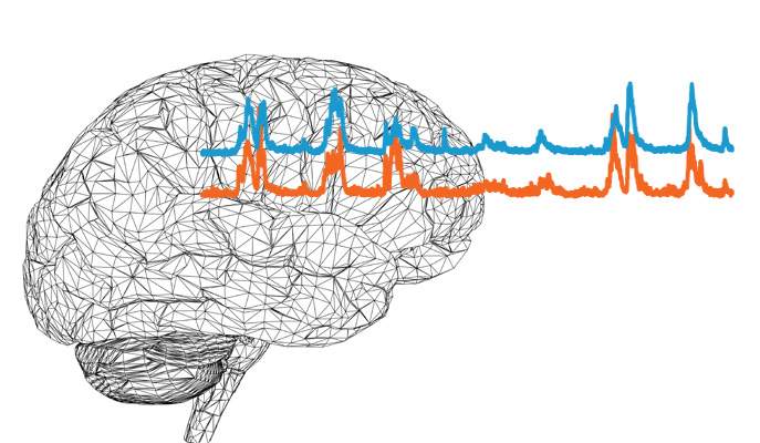
You would expect, then, that the pattern of activity generated in the brain cells would repeat itself when the cells respond a second or third – or fiftieth – time to identical stimuli. For example, a small response would be generated each time you looked at a particular chair. But this is not the case – the response tends to vary from trial to trial. So the next time you view the same chair, it could be that a large response would be produced in the brain cells.
Although different combinations of brain cells join the response each time, surprisingly, the cells that respond are synchronized – they respond to the sensory stimulus in the same way. The variability of the response in individual cells therefore cannot be accounted for simply by averaging out the responses of multiple cells.
What, exactly, is responsible for generating such “synchronized variability”?
Which camp do you belong to?
This question has been a matter of debate for decades, with scientists divided into two main camps. One camp believes that the variability and synchrony originate from signals coming from the part of the brain called the thalamus, which is responsible for routing most of the sensory stimulation the body detects to cells in the cerebral cortex, the part of the brain involved in processing and interpreting the sensory information.
The second group suggests that the variation in the observed responses and the synchrony are produced when signals are being relayed between a network of brain cells within the cortex region after having arrived from the thalamus.
Prof. Ilan Lampl, of the Weizmann Institute of Science’s Neurobiology Department, and his team, including postdoctoral fellow Dr. Katayun Cohen-Kashi Malina, former PhD student Boaz Mohar and PhD student Akiva N. Rappaport, finally shed light – literally – on the matter, placing them in the “cortex camp.”

Solving the debate
Brain cells within the cortex receive signals from, and send signals to, many other cells via junctions called synapses. The scientists devised a unique experiment in which they placed electrodes within cells in the cortices of mice and recorded the electrical current produced by the direct synaptic inputs. Because they set the experiment up to record the activity of nearby pairs of cells – as opposed to individual cells – they were able to test for and unveil the origin of synchrony.
To find whether or not the variability and synchrony originate from signals coming from the thalamus, the scientists employed a technique called optogenetics, which allowed them switch cellular activity in the cortex of the transgenic mice “on” or “off” using light: When they illuminated the cortex, the cortical cells were completely inhibited and therefore could not pass sensory information to others. Any electrical current detected with the electrodes in the cortex cells when these cells were inhibited would necessarily have been routed from the thalamus, as its activity was not affected by the light.
Lampl: “We were able to construe the recordings from the thalamus according to two possible scenarios: If two cells in the cortex are activated together upon receiving signals from the thalamus, it means that variability and synchrony originate from the thalamus. If, on the other hand, the cells are activated independently of one another, then we can deduce that the variability and synchrony originate from signals being relayed between cells within the cortex.” Two sets of recordings were conducted, one without sensory stimulation, the second with stimulation of the animals’ whiskers.
The team’s findings, published in Nature Communications, show that when inhibited brain cell activity in the cortex only allowed signals to propagate from the thalamus, the response of cortex cells was less variable and synchronized from trial to trial for both conditions. This provides evidence that it is, indeed, the signals that are relayed between cells within the cortex that are responsible for the observed variability and synchronization rather than those originating from the thalamus.
Is cell synchrony “good” or “bad?”
Lampl: “At the moment, the generation of cell variability and synchrony is an observation – a hallmark of physiology. We don’t know if it is a ‘good’ or ‘bad’ thing that many cells act together.” Understanding the origins and function of cell variability and synchrony could yield useful insights into information processing in the cortex and perception, not only for the tactile system that was studied but also for the visual and auditory systems, which have obtained similar findings.
Prof. Ilan Lampl's research is supported by the Henry Chanoch Krenter Institute for Biomedical Imaging and Genomics; the Marianne Manoville Beck Laboratory for Research in Neurobiology in Honor of her Parents; and the Adelis Foundation.