עיתונאיות ועיתונאים, הירשמו כאן להודעות לעיתונות שלנו
הירשמו לניוזלטר החודשי שלנו:

Some 100 years after the discovery of enzymes, most research on their activities still gets conducted in “test tubes” of various sorts. How well do these test-tube models explain what happens inside living cells? Prof Gideon Schreiber and his group in the Biomolecular Sciences Department of the Weizmann Institute of Science devised, for the first time, experiments to track the efficiency of enzyme reactions within living cells. Their findings, which recently appeared in the Journal of Biological Chemistry, suggest that the catalytic process that takes place within the cell may be chemically identical to that in the test tube – but it may be less efficient in its natural environment. The explanation for this may have implications for drug design, as well as for much of biochemical research.
Test-tube experiments, explains Schreiber, are useful because looking at the undisturbed interaction between a purified enzyme – the catalytic protein – and its substrate – the protein or molecule it binds to and alters – can reveal the exact nature of the mechanism. The enzyme efficiency of these reactions have also been exhaustively studied, but scientists have long suspected that in the crowded, complex, hectic environment of the cell, these reactions may work at a different pace than in the test-tube solution.
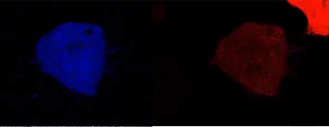
To track the binding of an enzyme to a substrate and the substrate’s subsequent alteration – the product – inside a living cell, the researchers, Agnes Zotter of Schrieber’s group and Felix Bäuerle of the Max Planck Institute for Dynamics and Self-Organization, had to go back to the drawing board and develop methods that would enable them to quantify the process. They began by inserting genes for an enzyme normally found in bacteria into human cells in a lab dish. These genes were engineered to create enzymes that glowed red under the microscope, and the researchers developed an algorithm to count enzymes by the intensity of the light.
The substrate they used glowed light green in its original state and bright blue when it was turned into the product. This substrate was inserted into each cell individually through a technique known as microinjection.
The team produced Technicolor videos for their cells, tracking in real time the amounts of enzyme and product. From these videos, they developed algorithms to plot the rates at which these steps occurred, and they tested these algorithms against more conventional methods to verify them.
Until now, we have not understood what this means once the molecule is inside the cell
The researchers noted that the amount of enzyme produced varied from cell to cell. What was puzzling was the fact that the efficiency of the reaction dropped as the amount of enzyme increased. The efficiency, explains Schreiber, is similar to that of a factory: It is based on the total amount of final product produced each second or hour, divided by the number of machines. Further simulations suggested that the strange result was due to a shortage of substrate; but the researchers’ measurements showed plenty of substrate in the cell, so they knew this could not be the case.
Further simulations suggested an alternate explanation: Much of the substrate gets tied up in other ways, so that it is not free to react with the enzyme. This was then experimentally tested by using focused light to “bleach” the substrate in a small area of the cell and observe how fast the dark spot was refilled by the surrounding molecules. These small molecules, according to predictions, should quickly diffuse through the cell, but instead they were relatively sluggish.
The researchers then asked what was holding up the substrate. One way to understand the biochemistry of a protein in a cell is to ask where it lies on the scale of water-loving to water-hating. The substrate turned out to lie toward the water-hating, or hydrophobic, end of the scale. This property made it somewhat sticky – apt to weakly bind to a number of other proteins in the cell. Even weak binding makes the substrate “invisible” to the target enzyme, until it frees itself once again.
Schreiber and his team then compared the hydrophobicity of the substrate to that of drug molecules that are often used today, finding that the substrate fell within the accepted range for medical use. “Small molecules that enter the cell, to inhibit an enzyme, for example, need to be hydrophobic enough to pass through its fatty, hydrophobic outer membrane – though not so hydrophobic that they get stuck there,” says Schreiber. “But until now, we have not understood what this means once the molecule is inside the cell.
“One would think that adding more enzymes – the ‘machinery’ – would increase the output, but the model we obtained shows that the amount of the enzyme is almost irrelevant. You can add more ‘machines’ and get the same rate of production. The substrate is what limits the efficiency of the process, but the amount of free substrate may be much less than the actual amount of that substrate in the cell,” says Schreiber.

And there may be other limiting factors: A drug, for example, which must enter from outside the cell, may be limited by membrane “gateways.” On top of this, many processes in the cell involve the activity of multiple enzymes, so that the product of one reaction becomes the substrate of the next. “We don’t yet know how the efficiency changes at each step,” he adds.
This is the first time that reaction rates of an enzyme have been tested inside living cells. The study’s surprising results may have implications not only for biomedical research, but for biochemical, test-tube based research in general.
Enzyme activity in a cell: Enzymes are red, the final product, which is fully converted within several minutes, is blue
Prof. Gideon Schreiber's research is supported by the Dana and Yossie Hollander Center for Structural Proteomics, which he heads; and the Henry Krenter Institute for Biomedical Imaging and Genomics.