עיתונאיות ועיתונאים, הירשמו כאן להודעות לעיתונות שלנו
הירשמו לניוזלטר החודשי שלנו:

How do bones know when they have reached their “destination” – that is, how do they decide they are at the right length and the right strength? Do they have a written map or internal GPS navigation system, or do they have to ask the “locals” for directions? Two new studies at the Weizmann Institute of Science suggest that for the bones, nearby muscles play an unsuspected role in bone healing, as well as in keeping the skeleton healthy and well aligned, on track to the proper “address.”
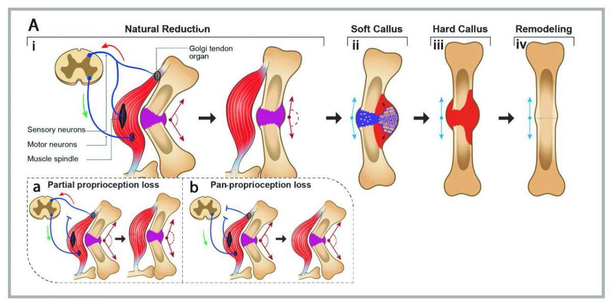
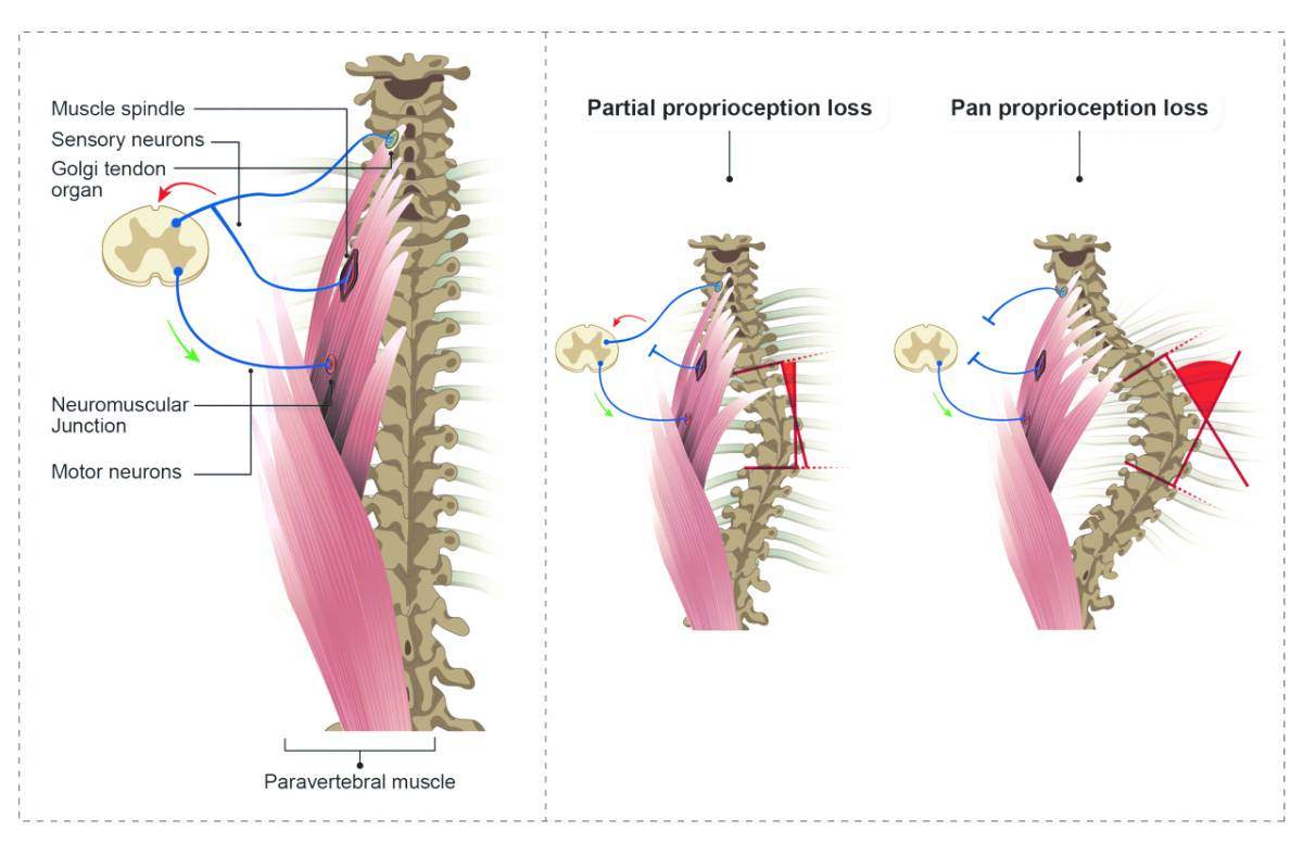
According to the studies, these directions come from a sensory system based in the muscles. To understand this system, you can close your eyes and touch a finger to your nose. The sense that allows you to do this – that lets you know, without looking, what your arms and legs are doing – is called proprioception (from the Latin, proprius, “belonging to me”). Proprioception is sometimes called the sixth sense. Unlike our other five senses, proprioception perceives not the external world, but rather the internal one: sensing our own bodies and the shifting placement of our limbs. Two “mechanical” sensors within the muscles are crucial to this sense: These are the “muscle spindles” found in the muscle fibers, which respond to changes in length; and the “Golgi tendon organs” found where the muscles connect to the tendons, which gauge the amount of contraction in the muscle. Prof. Elazar Zelzer’s research in the Weizmann Institute of Science’s Molecular Genetics Department suggests that these sensors – for extension and contraction – actually direct the development of the skeletal system.

The first hints of this arose in research conducted several years ago in his lab. He and his group had discovered that the broken bones of newborn mice “know” how to realign themselves fairly quickly, even without medical intervention. This finding contradicted the common assumption that broken bones first join at an angle and only then undergo “remodeling” with new bone to straighten out. Realizing that this alignment process was tied to the activity of the muscles, the researchers injected Botox into the mice’s muscles, thus paralyzing them, and found that broken bones, indeed, set at an angle without the muscles’ activity.
Their spines were taking on curves for no apparent reason
In the present study, which was recently published in Cell Reports, Zelzer and his group zeroed in on two genes required for the activity of the muscles’ sensory receptors – Runx3 and Erg3. This research was led by Dr. Ronen Blecher, an orthopedic surgeon in Assaf Harofe Medical Center, who was conducting PhD research in Zelzer’s lab. Together, they found that without these genes, the bone realignment did not take place. That is, the “sixth sense” plays a role in maintaining the health of the skeletal system.

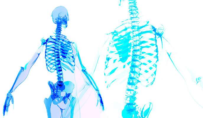
So far, the findings fit the group’s predictions; but Blecher, with his experience in interpreting medical X rays, noticed something that had not been foreseen: The mice lacking proprioception genes were developing the symptoms of scoliosis. Their spines were taking on curves for no apparent reason, and with no other apparent skeletal pathologies. Something similar happens in human cases of scoliosis in young people. These findings, which recently appeared in Developmental Cell, point, for the first time, to the involvement of proprioception in preserving correct spinal alignment, and in the progression of scoliosis, which had long been thought to be a disease of the bones. To achieve these findings, Zelzer and his team worked with the lab of Prof. Yoram Groner, who for decades has investigated the Runx family of genes, including their connection to proprioception.
“Every textbook will tell you that muscles are for movement and bones for structure, but this is something of a reversal of roles. The muscles determine, to a large extent, the structure of the bones,” says Zelzer. “The information on skeletal formation is not necessarily found only in genetic ‘maps’ in the bone, but rather in the interactions between muscle and bone, and the mechanical play of forces between them.”

Zelzer points out that it was the multidisciplinary nature of the study that led them to their surprising findings: “I am a developmental biologist, so I have a good understanding of how the skeleton forms. But it took collaboration with a physician, who could not only recognize pathologies in X rays, but who comes to the research with a different viewpoint. When we broaden our outlook – from just bones or tendons to whole systems, for example – we can obtain new findings that we would not otherwise have been able to observe.”
Prof. Elazar Zelzer's research is supported by the David and Fela Shapell Family Center for Genetic Disorders Research, which he heads; the David and Fela Shapell Family Foundation INCPM Fund for Preclinical Studies; the Weizmann-University of Manchester Collaborative Projects Program; and the estate of Bernard Bishin for the WIS-Clalit Program.